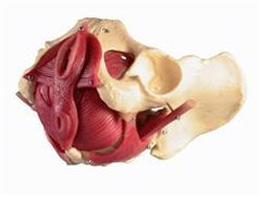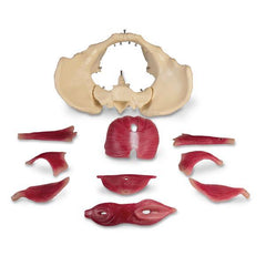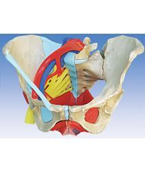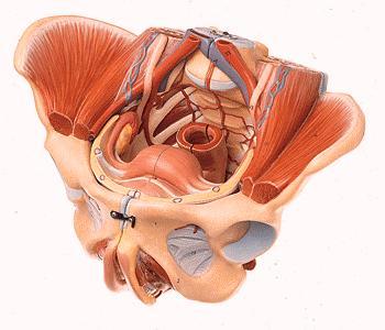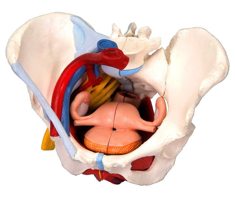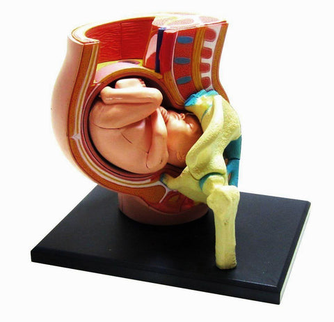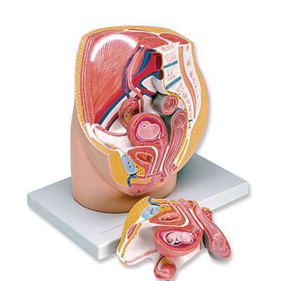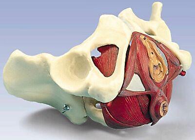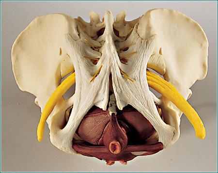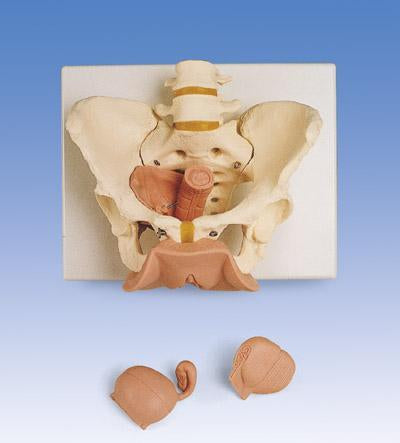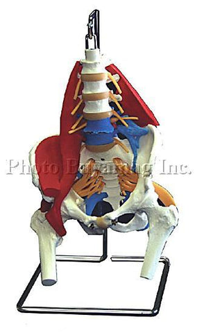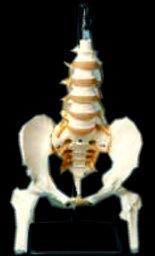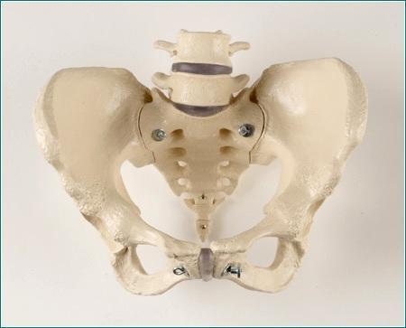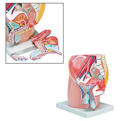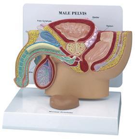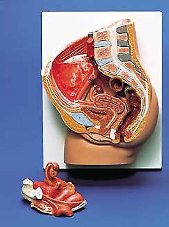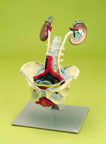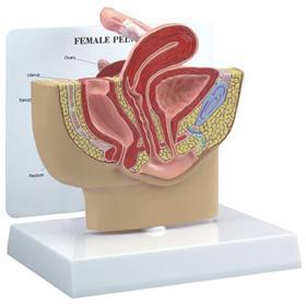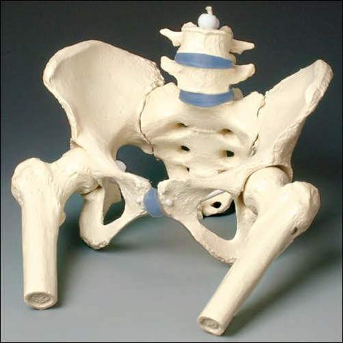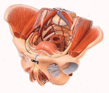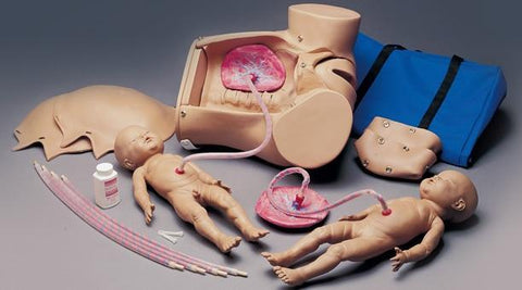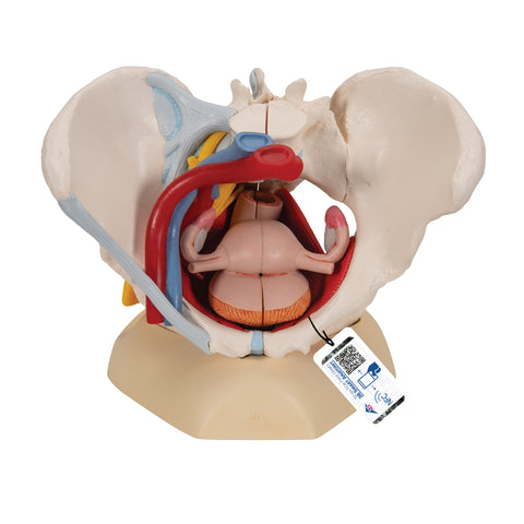
Female pelvis anatomical model represents the pelvic floor in its layers. The following muscles are represented and can be removed.
This Anatomical Model is used by medical schools in student's educational programs for easy to learn and understand Human Anatomy, Pathology and disease prevention. In doctor's and chiropractors offices for patient's education. Also used for legal presentations. Weight: 5 lbs.
Features
- Left and right obturator internus muscle
- Left and right piriformis muscle
- Left and right coccygeus muscle
- Pelvic diaphragm (levator ani muscle consisting of puborectalis muscle, pubococcygeus muscle, and iliococcygeus muscle)
- Urogenital diaphragm (consisting of the deep transverse muscle and the superficial transverse muscle of the perineum, and the ischiocavernosus muscle)
- Sphincters of the urogenital and digestive tract (consisting of external anal sphincter, urethral sphincter, and bulbospongiosus muscle)
- Together with the hip bones and the sacrum, the model consists of 12 parts total. Muscles are fixed with pins, allowing them to be removed for demonstration of the layers. 10-5/8" x 7" x 6-11/16".

