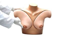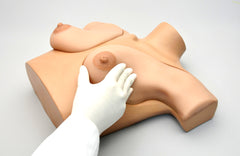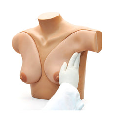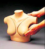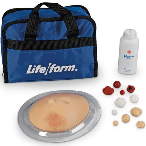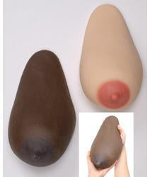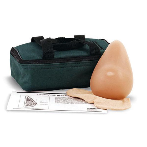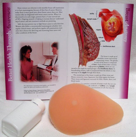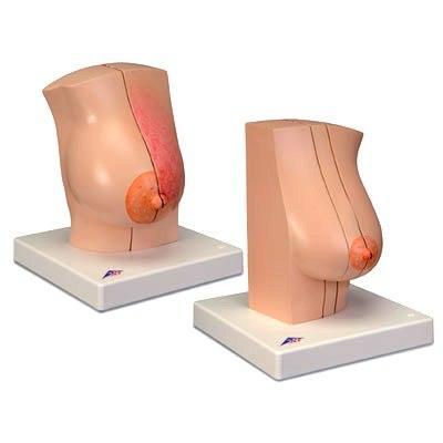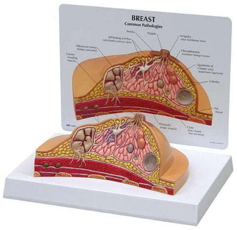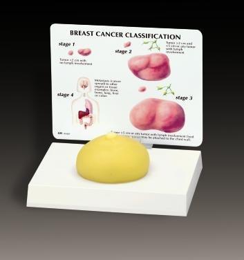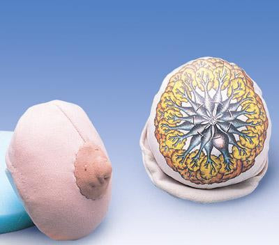
Breast Self Examination Simulator Model Manikin
Buyamag INC
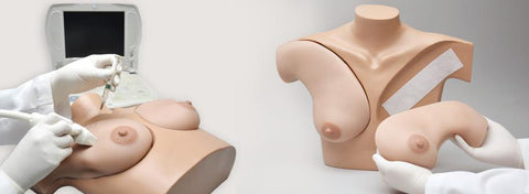
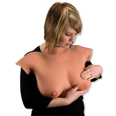
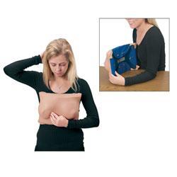
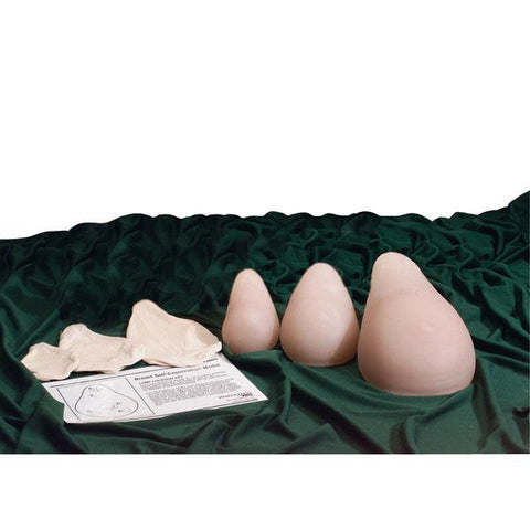
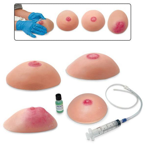
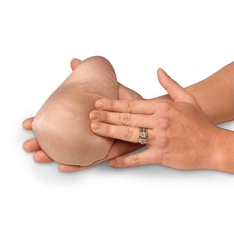
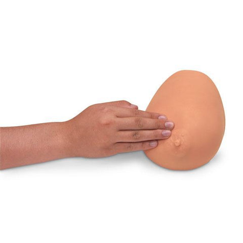
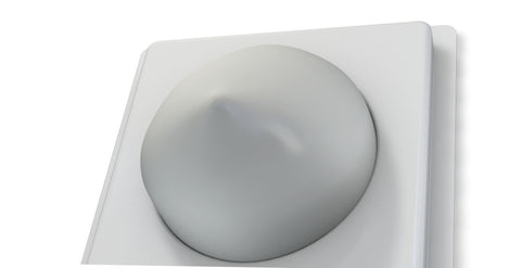
Breast self examination simulator model developed to assist women and health professionals in teaching the processes and skills required to perform both: self-examinations and clinical Identification of pathology conditions.
Left and right breasts firm & easy attaches with Velcro

It is not possible to determine the exact nature of any tumor strictly by touch. Although many breast lumps are not cancerous, any lump should be examined by a health professional. With early detection - through breast self - examination, clinical exams by a healthcare professional, mammography - the chances of surviving breast cancer increase dramatically.
All women should perform monthly breast self- exam. Women after 40 and older should have a clinical breast exam every year.
Simulator received top marks at a recent global health meeting. Healthcare professionals were elated with its realism, durability, materials and texture.
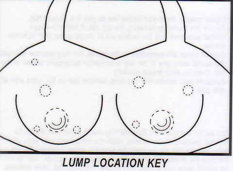
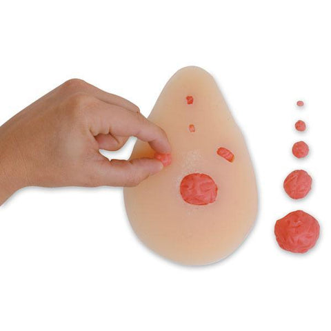
- Left breast contains four masses in the breast tissue and two masses in the axilla region. The masses range in size from 14 to 19 mm, and their depths range from 6 to 16 mm beneath the surface.
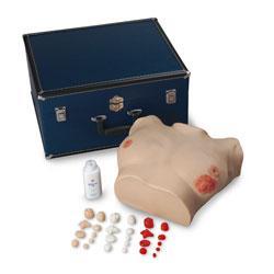
- Right breast contains enlarged lymph node, fibroadenoma, fibrocystic mass and fluid-filled cyst (ranging from 12 to 24 mm)
- Breasts are attached to a manikin torso and can be easily removed and reassembled
- Simulator can be used in either the upright or reclining position
- Medium skin tone standard
- Optional light or dark skin tone
- Soft carrying bag
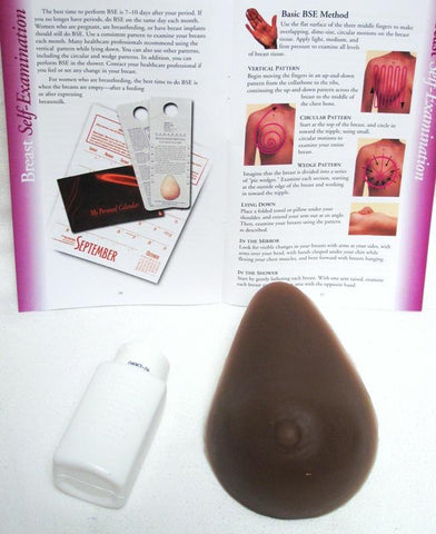
- User Guide
- Realistic texture and look
- Durable

