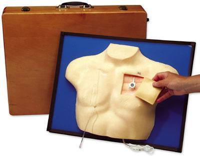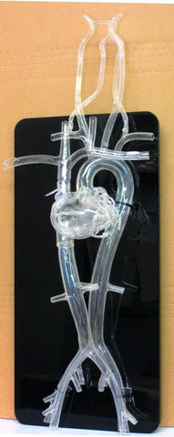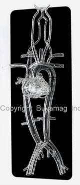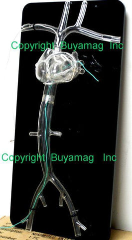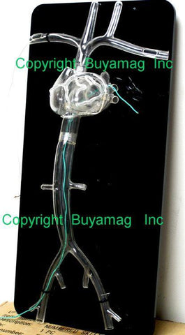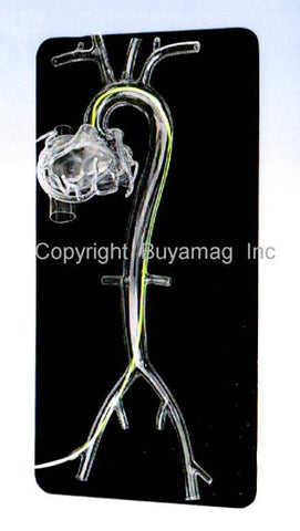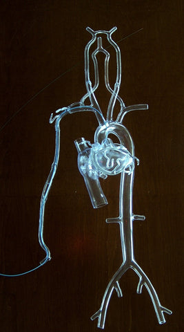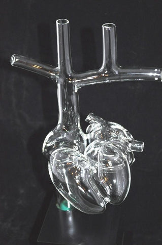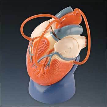
E.P. Heart Practicing Catheter, Lead, Electrophysiology Heart Deluxe
Buyamag INC
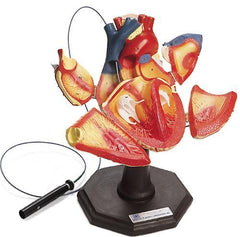
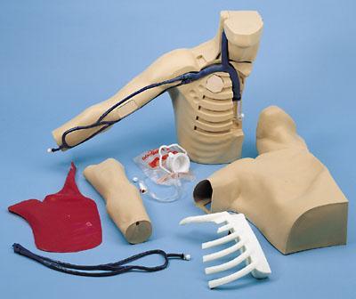
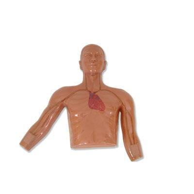
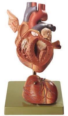
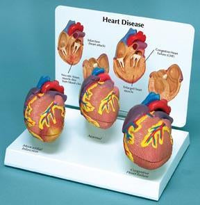
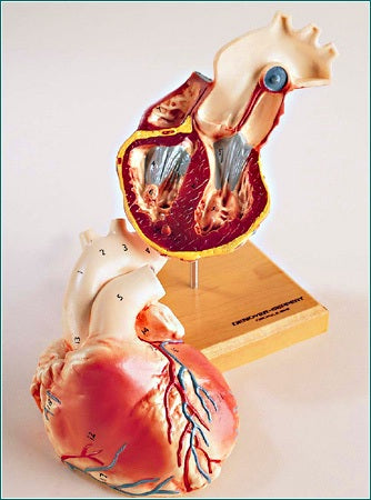
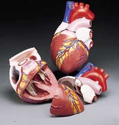
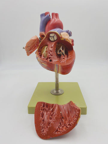
Four hinged doors lead to the Heart Chambers of this twice life-size Heart Model, complete with a display stand. Additional openings which allow for catheter and lead placement, include: Brachiocephalic Vein, left Subclavian Vein, superior Vena Cava, Aorta, Aortic Valve, inferior Vena Carva, orifice of the Coronary Sinus, and Atrial Septal defect. Actual pacing leads can be positioned in the right Atrial Appendage, right ventricle orifice of the coronary sinus, through the ASD, and in the left ventricle. 60 coded structures are identified in the 8 page instructional guideExcellent for teaching purposes with respect to Pacing and Electrophysiology. Designed as an excellent practicing, educational, teaching and patient presentation tool. Real-Like Colors, detailed hand painted outside inside. Four Chambers on hinges, easy open or closed. Highly detailed articulated model makes easy understand the treatments procedures, anatomy, function and pathology of the human heart This highly detailed Heart Model is excellent for medical schools and education programs to help students understand the heart, diseases, function and anatomy. Demonstrations in doctors offices and legal presentations. Catheter not included.

